
Overview
Whole body scanning by computed tomography (CT) is being vigorously marketed as a means of screening for early signs of illness in people who have no symptoms or disease risk factors. The claimed benefit is that diseases such as cancer can be treated more successfully if they are detected in their early stages.
CT scanning has a valuable role to play in investigating suspected problem organs, or in circumstances where there is a high probability of illness. The new service that is being promoted, however, is the use of CT scanning as a broad screening tool.
There are significant risks that outweigh the benefits associated with the whole body scanning of otherwise healthy people. After considering the available information, the Authority and the Radiation Advisory Council (an expert body established to advise the Authority) are of the view that the procedure is inappropriate for general diagnosis of healthy individuals. The Authority is taking strong steps to ensure that people who choose to go ahead with a whole body scan are made fully aware of the risks involved.
This publication gives an overview of whole body scanning and discusses its risks and limitations. General guidance is also provided on when to have a CT scan and what factors to consider when making this decision.
CT scans and how they work
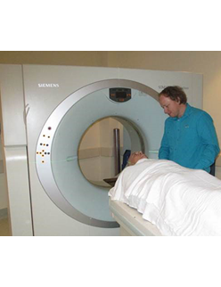
Computed tomography (CT) has only been widely available for the past 25 years and was made possible by the coupling of x-ray equipment with computer analysis. In traditional medical diagnostic x-ray apparatus, low doses of x-rays are passed through tissues in the body onto a photographic plate to produce a two-dimensional image.
In CT, the x-rays are targeted through the particular internal part of the body of interest at many hundreds of angles for every cross sectional slice. The information from these x-rays after they have passed through the body is analysed by a computer, which then creates a series of detailed cross-sectional images.
CT imaging is most useful in the examination of specific target organs in the body using a narrow beam of x-rays, often with the use of a contrast medium to enhance the detail seen in an organ scan. Other imaging technologies such as conventional x-ray and ultrasound usually only capture a limited number of internal features such as soft tissue, bone or blood vessels. CT is good at capturing details of all three.
CT is the technique of choice for examinations of the pelvis, chest, abdomen, brain and spine, and is particularly helpful in the diagnosis of lung, liver and pancreatic cancer, but not for ovarian or prostate cancer.
CT is also used to measure the size and precise location of tumours; to evaluate the extent of cancer spread; and as an aid to guiding biopsies and radiation treatments. Other uses include the evaluation of abscesses and other inflammations; for detecting abdominal aneurisms and other vascular problems; and the examination of kidney stones.
Risk associated with the large radiation doses from whole body CT scans
The 2000 report of the United Nations Scientific Committee on the Effects of Atomic Radiation (UNSCEAR) determined that CT imaging administers the largest medical radiation dose to the population. It is estimated that CT now accounts for 40% of the total dose of radiation from all types of diagnostic x-ray examinations in the United Kingdom, despite amounting to only 6% of the total number of such examinations.
This represents a doubling in the radiation dose from CT in the last 10 years or so, mainly due to the rapid increase in the number of examinations. CT examinations are high-dose procedures which, when used correctly, are of substantial benefit to the population. New technologies and new techniques allow CT to be used for a wider range of procedures, and the increase in the radiation dose will most likely continue or even escalate.
The International Committee on Radiological Protection (ICRP), in its recommendations (Publication No. 60, 1990) established three main principles for radiation dose reduction. These are:
- justification of the benefit of a radiation procedure as measured against possible detriment
- minimisation of the dose to the lowest possible level to be effective for a procedure
- limiting the amount of radiation to which the patient is exposed.
In the case of whole body CT scanning it is difficult to justify medical examinations that involve large radiation doses which may be of little benefit to the patient. Whole body scanning currently offered in Sydney imposes large radiation doses on clients in the range of 4 to 24 millisieverts (mSv) per scan. In comparison, a chest x-ray gives an average dose of 0.04 mSv.
The Department of Health has advised that a whole body CT scan with an effective dose of 10 mSv may be associated with an increase in the possibility of cancer of approximately 1 in 2000. This cancer risk generally outweighs the potential health benefits for healthy people under the age of 50.
Limitations of the procedure
Although CT imaging may be useful as an investigative tool for some potentially fatal diseases, general whole body CT scanning in otherwise healthy individuals can also have negative consequences. For example, patients who receive a clean bill of health after a whole body CT scan may be left with a false sense of security about their health, discouraging them from adopting healthier lifestyles or from having regular medical check ups or other more appropriate screening tests.
On the other hand, people whose scans produce suspicious findings may be subjected to expensive, invasive and sometimes unnecessary follow-up medical procedures. In healthy people, about 80% of abnormalities detected on CT studies are relatively harmless findings such as benign nodules, non-cancerous tumours, and scar tissue from past infections.
In addition, what constitutes a 'normal' scan differs with age, and the high level of expertise necessary to recognise these distinctions may not always be available.
Worldwide, CT scanning has not been fully evaluated as a general screening tool, so there are no established protocols, and professional and government organisations do not endorse whole body scans. The Royal Australian and New Zealand College of Radiologists issued a press statement in June 2002 warning consumers of the limitations of the practice.
Concerns expressed in the United States
In response to concerns expressed by the US Food and Drug Administration (FDA), the National Council on Radiological Protection and Measurement is developing a set of guidelines on the management of patient dose in CT scanning. The FDA is developing a tutorial on the dangers associated with CT scanning. This will be available on their website when it is completed.
The American College of Radiology issued a statement on 27 September 2000 in which it proposed that there is not sufficient evidence to justify whole body CT screening in people with no symptoms or family history of illness. The College also expressed concerns that the procedure would lead to findings that will not be of benefit to the person's health but would rather result in increased anxiety, unnecessary follow-up examinations and treatments, and wasted expense.
When to have a CT scan
Even when the quality of whole body CT scanning is high, the procedure is not a substitute for other screening tests, such as periodic mammography for breast cancer; a pap smear for cervical cancer; bone densitometry for osteoporosis; blood tests for prostate cancer and diabetes; and tests for heart disease and blood pressure. Although CT imaging is valuable for detecting colon cancer, it is not a substitute for the more traditional techniques of sigmoidoscopy and colonoscopy. It is not yet known how effective CT scanning is in screening for lung cancer.
There is no doubt that CT scans are advisable when a medical evaluation indicates that it may provide helpful diagnostic information. Such a medical evaluation should be performed by an independent doctor taking into consideration symptoms as well as disease risk factors, such as blood cholesterol and glucose levels; blood pressure; smoking and exercise habits; weight and diet; occupation; and the medical history of family members.
However, because CT scanning has not been widely studied as a general screening tool, it should be reserved for particular cases where the symptoms and risk factors indicate that it is advisable.
What should be considered when undergoing a whole body CT scan
- The scan should only be undertaken as part of a comprehensive evaluation, including a physical examination, medical history and other tests.
- You should not undergo such a test without a written request from an independent medical practitioner.
- Whole body scanning is not recommended for people under 50 years as they are more at risk of developing cancers as a result of this procedure.
- You should understand the risks involved and the scale of the radiation dose you will receive. These should be clearly explained to you by a medical practitioner.
- You should be asked to give your informed consent in writing prior to undergoing the whole body CT scanning procedure.
The Radiation Advisory Council provides the following guidelines when undergoing a CT body scan:
- Choose a radiologist who specialises in CT imaging . The radiologist should also communicate with your medical practitioner.
- Choose a facility that is affiliated with a major health centre. These providers are more likely to have high standards in terms of staff and equipment.
- Choose a practice with only the most up-to-date and best equipment . The best equipment is the multi-detector CT with three dimensional reconstructions. Other CT scanners that are also appropriate include the spiral or helical scanner and electron beam CT. Patients should be aware that other specific procedures are also required in the case of virtual colonoscopy and certain other applications such as evaluation of the coronary arteries for calcium deposits.
- In some examinations such as for abdominal (but not chest) scans, a contrast medium should also be used. This helps to differentiate between normal and abnormal tissues. Without the use of a contrast medium, small lesions of the liver, kidneys and pancreas may be missed, and any lesions that are detected may not be fully characterised. Discuss with your referring doctor if a contrast medium is required.
- Appropriate patient preparation should be performed for the organ being investigated (for example clearing of bowel contents for best results in virtual endoscopy). Check with your referring doctor as to what preparation is required.
Further Information
- United Nations Scientific Committee on the Effects of Atomic Radiation, Report 92-1-142238-8 Sources and Effects of Ionizing Radiation, Volume 1 Sources, 2000.
- D. Hart, M. C. Hillier, B. F. Wall, et al., Report United Kingdom National Radiological Protection Board – R289 Doses to Patients from Medical X-ray Examinations in the UK – 1995 Review, 1996.
- International Commission on Radiological Protection, Publication No. 60 1990 Recommendations of the International Commission on Radiological Protection, 1991.
- International Commission on Radiological Protection, draft publication Diagnostic Reference Levels in Medical Imaging, February 2001.
- International Commission on Radiological Protection, Publication No. 87 Managing Patient Dose in Computed Tomography, October 2000.
- Australian Radiation Protection and Nuclear Safety Agency, Radiation Protection Series No. 1 – Recommendations for Limiting Exposure to Ionizing Radiation (1995) (Guidance Note [NOHSC:3022(1995)]) and National Standard for Limiting Occupational Exposure to Ionizing Radiation [NOHSC:1013(1995)] March 2002.
- Johns Hopkins University School of Medicine, Baltimore, Maryland, United States of America, Health After 50 Program: A Full Body Scan for Everyone? April 2002.